Ketone Bodies and Glucose: Competitors or Team Players?
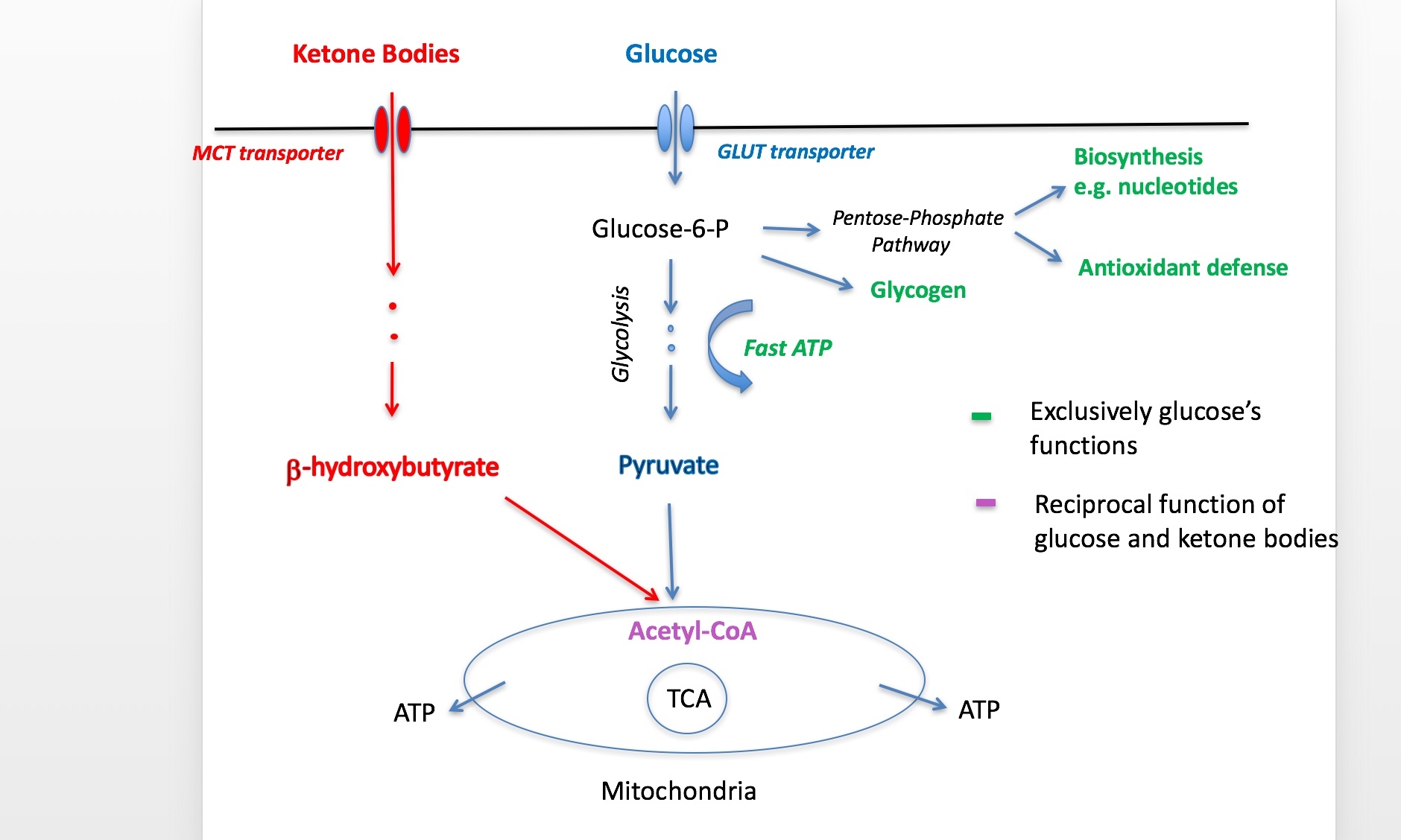
It is our recent proposition that ketone bodies can spare glucose to fulfill its energy-supplying and exclusive neuroprotective functions. In normal physiological conditions glucose still can be effective in both. In the conditions of pathological glucose hypo-utilization as it seen in all neurodegenerative diseases, these functions suffer. Importantly, it results in oxidative stress and insufficient energy reserves in the brain, partly due to inadequately low brain glycogen stores. This explains why during brain pathologies, on ketogenic diets, ketone bodies fulfill their glucose sparing function allowing sufficient amounts of glucose be allocated toward its neuroprotective role.

Pyruvate Supplementation Increases Exploratory Activity and Brain Energy Reserves in Young and Middle-Aged Mice
Metabolic assessment of brain glycogen levels. (A) Group means and individual values for cortical glycogen content in the enzymatic assay in young adult wild-type mice at 6 months of age after been on dietary intervention for 3 months by the time of euthanasia. PYR group has higher brain glycogen levels (*p < 0.05, t-test). (B) PAS staining for brain glycogen in 13-month-old wild-type mice been on the dietary intervention for 7 months. Data are given as relative optic density (OD%) with 0 corresponding to pure white and 100% to pure black. DG = dentate gyrus, molecular layer, CA1 = all layers of this subregion, cc = alveus + corpus callosum, and Ctx = all layers of visual cortex. PYR group has higher PAS staining intensity, *p < 0.05, **p < 0.01 (t-test). (C,D) Representative PAS-stained sections through the mid-hippocampus after conversion to grayscale. Scale bar = 500 μm. C = STD group and D = PYR group.
Source: Koivisto H, Leinonen H, Puurula M, Hafez HS, Barrera GA, Stridh MH, Waagepetersen HS, Tiainen M, Soininen P, Zilberter Y and Tanila H (2016) Chronic Pyruvate Supplementation Increases Exploratory Activity and Brain Energy Reserves in Young and Middle-Aged Mice. Front. Aging Neurosci. 8:41. doi: 10.3389/fnagi.2016.00041